IMPLANTOLOGY
Bone Grafts
When bone deficiency is identified, a bone graft may be required to prepare the implant site and ensure the long-term stability of the future implant.
Schedule an initial consultation →
When bone deficiency is identified, a bone graft may be required to prepare the implant site and ensure the long-term stability of the future implant.
Schedule an initial consultation →Bone grafting makes it possible to place a dental implant even when the available bone volume is insufficient.
At the 47VH clinic, we master the full range of bone reconstruction techniques, including autogenous bone grafting, biomaterials, guided bone regeneration, and sinus lift procedures. After analyzing your 3D radiological assessment, we define the most appropriate solution for your situation together, with the fewest possible surgical procedures.
A sufficient amount of bone is essential for placing a dental implant and ensuring its long-term stability.
When bone volume is insufficient, the missing bone must be reconstructed to create a stable foundation for the future implant.
In such cases, a bone graft is performed. Depending on your clinical situation, it may be carried out before implant placement (pre-implant bone grafting) or simultaneously with implant placement (peri-implant bone grafting).
We use a wide range of materials and techniques for bone reconstruction. The choice depends on the selected surgical approach as well as the patient's preferences and clinical needs.
In this approach, bone is harvested directly from the patient, usually from the mandible. Because it requires a second surgical site, the procedure is more invasive and generally increases the duration of surgery.
Because two surgical sites are involved, post-operative discomfort is often more significant. However, this type of graft generally provides the best biological properties, as the bone is completely biocompatible and originates from the patient.
In this approach, bone from a human donor is used. It is typically obtained from donated femoral heads and is carefully processed and sterilized to ensure safety and eliminate the risk of disease transmission.
This processing reduces the graft’s osteogenic potential, but it simplifies the procedure: surgery is generally shorter than with an autogenous graft and requires only a single surgical site. However, outcomes may be somewhat less predictable depending on the clinical situation.

Bone biomaterials provide an effective alternative to autologous bone grafts. Their use can shorten surgical time and reduce morbidity, although the clinician must adapt the surgical technique to achieve predictable and successful outcomes.
Bone biomaterials are derived from different sources. They are generally divided into two main categories:
These bone substitutes most often come from bovine tissues. Laboratories treat them to eliminate any risk of contamination.
Used for more than 20 years and extensively documented in the scientific literature, these biomaterials allow for shorter procedures with predictable and reproducible outcomes. They are now considered the reference material for many bone augmentation procedures. At the 47VH clinic, we preferentially use premium biomaterials supported by high-quality scientific evidence.
Synthetic substitutes are manufactured through chemical processes. Their biological properties remain limited, and clinical outcomes are often less predictable. For this reason, we use them only rarely in our practice.
Growth factors are small proteins capable of accelerating biological processes — such as healing or bone formation.
Their use is, however, prohibited in Europe and many other countries. Although some protocols or biomaterials claim to incorporate them, current scientific evidence remains insufficient to support their routine clinical use.

In certain cases, we use shorter or narrower dental implants to avoid the need for bone grafting procedures.
Recent technological advances have led to the development of small-diameter and short implants with improved mechanical performance. However, they are not suitable for every clinical situation, and their use depends on a variety of anatomical, functional, and prosthetic factors.

In some cases, it is essential to rebuild the bone in height, width, or both, in order to allow the placement of dental implants.
Today, a wide range of effective techniques is available to restore deficient bone volume. The choice of method depends primarily on the type, extent, and location of the bone defect.
When bone loss is significant, bone reconstruction is generally performed before implant placement. A healing period of approximately 4 to 9 months is then required before the dental implant can be inserted.
When bone loss is moderate, bone reconstruction and implant placement can often be performed during the same surgical procedure, simplifying treatment and reducing the overall treatment time.
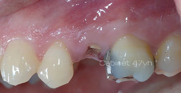
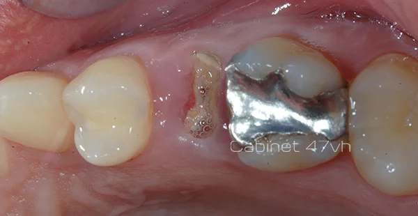
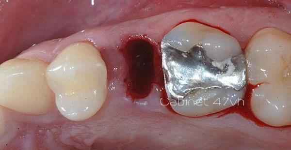
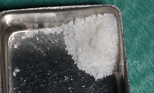
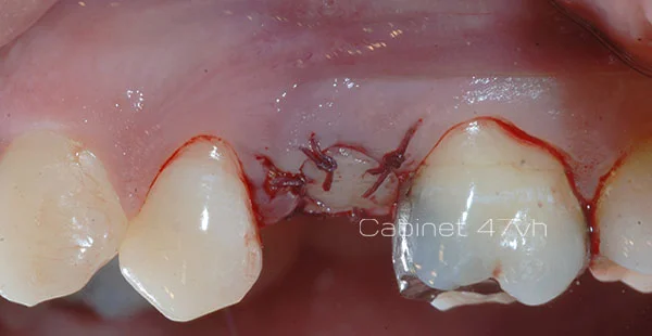
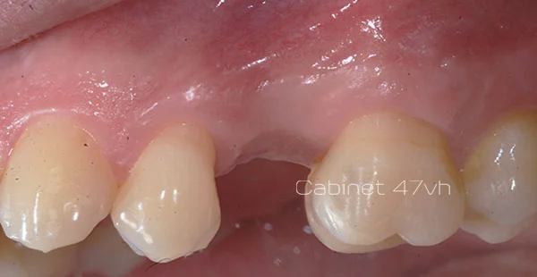
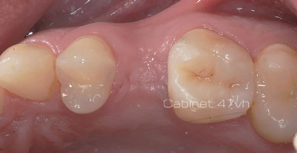
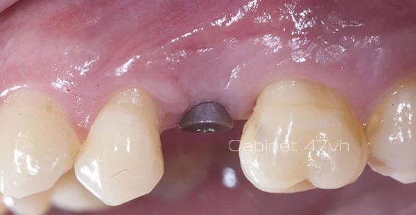
To minimize bone loss following tooth extraction, we place a bone biomaterial directly into the extraction socket during an atraumatic extraction and then protect it with a collagen matrix or a gum graft.
This approach preserves bone volume from the time of extraction, facilitates future dental implant placement, and, in most cases, helps avoid more complex bone grafting procedures.
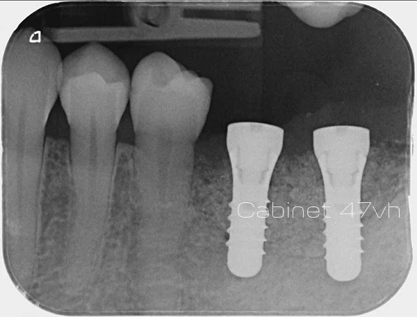
Extraction with alveolar preservation
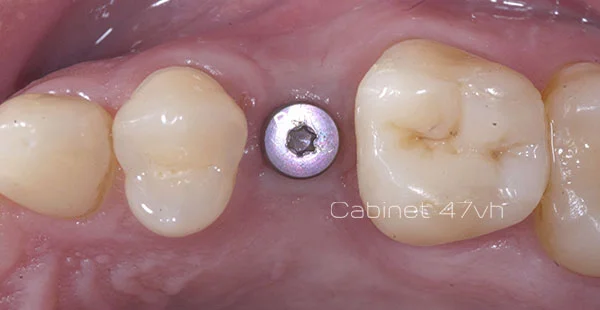
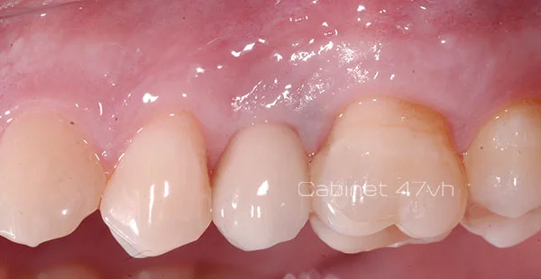
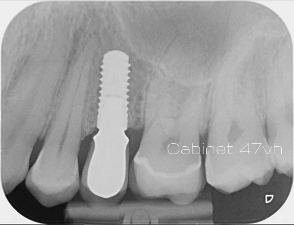
The maxillary sinuses are air-filled cavities located within the facial bones, above the premolars and molars of the upper jaw.
After the extraction of these teeth, the amount of bone available beneath the maxillary sinus is often insufficient for dental implant placement. In such cases, a sinus floor elevation procedure—commonly known as a sinus lift or sub-sinus bone graft—is performed to recreate adequate bone volume for implant placement.
A small lateral opening is created in the bone, and the sinus membrane is carefully elevated. A bone biomaterial is then placed beneath the membrane to maintain its new position and create a space where new bone can form.
Your own bone cells then gradually regenerate the missing bone. In most cases, sufficient new bone forms within approximately four months, allowing dental implant placement under favorable conditions.
We mainly use two approaches, depending on the amount of available bone:
We prefer this technique when bone loss is significant. Surgical access is gained through the lateral wall of the sinus, making it a reliable and highly predictable procedure. In most cases, implant placement is performed at a later stage, after adequate bone healing and consolidation.
We use this approach when bone resorption is moderate. Access is obtained directly through the alveolar ridge during implant site preparation. This minimally invasive technique is quick, associated with limited post-operative discomfort, and generally allows implant placement during the same surgical procedure.
We choose the technique best suited to your bone situation to ensure effective and durable reconstruction. The sinus lift thus makes it possible to place dental implants even in areas with significant posterior maxillary bone deficiency.
Lateral window sinus lift
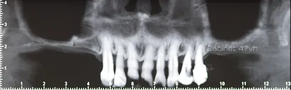
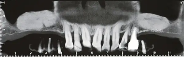
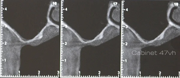
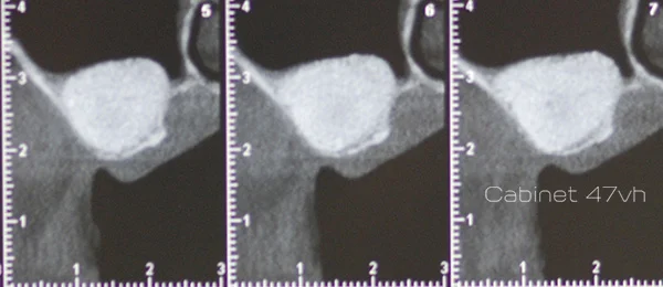
Guided bone regeneration stimulates the formation of new bone by creating a protected space that allows your own cells to regenerate the missing tissue naturally.
A bone biomaterial is placed, sometimes combined with autologous bone chips, and then stabilized with a synthetic or collagen membrane that creates a protected environment favorable to bone regeneration.
These techniques have evolved considerably in recent years, and many variations are now available. The choice of approach depends on the specific clinical needs of each patient.
Their implementation is technically demanding and requires an experienced surgeon. However, these techniques can regenerate substantial volumes of high-quality bone without the need for extensive bone harvesting from another part of the body.
In certain cases, dental implants can be placed at the same time as the bone regeneration procedure, reducing the number of surgeries and shortening the overall treatment time.
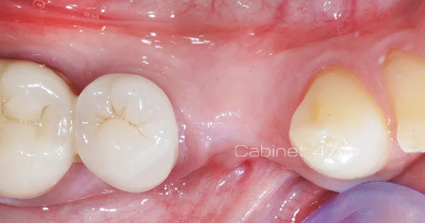
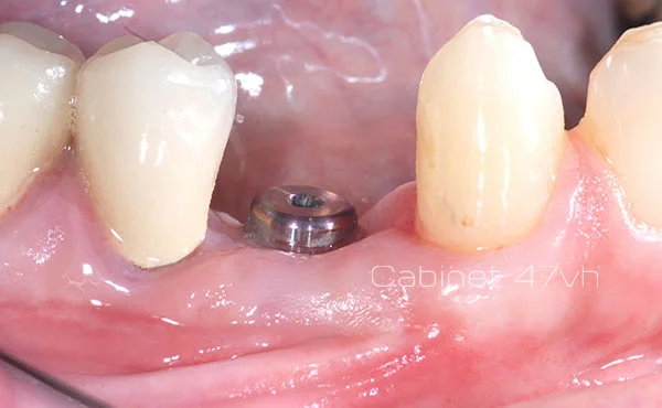
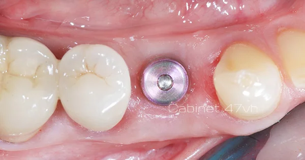
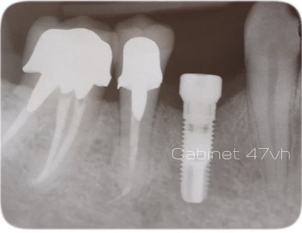
Simple horizontal guided bone regeneration
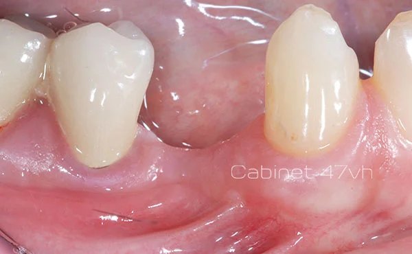
Advanced horizontal guided bone regeneration
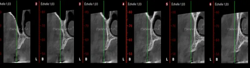
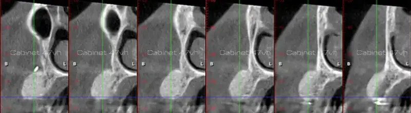
Vertical guided bone regeneration
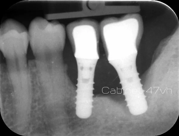
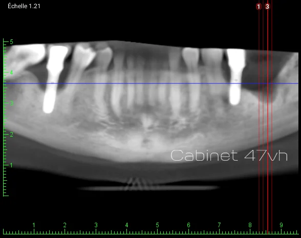
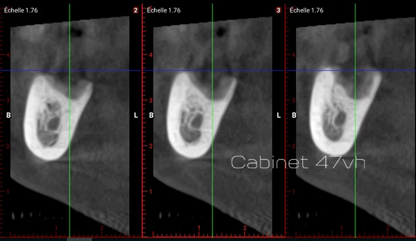
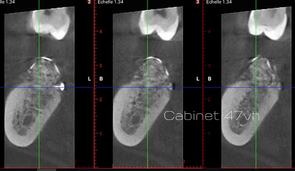
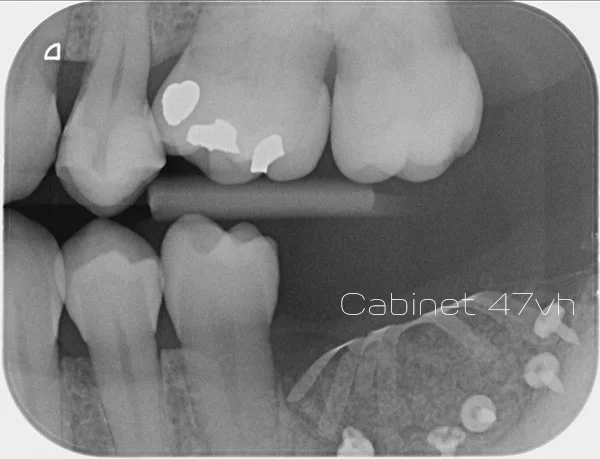
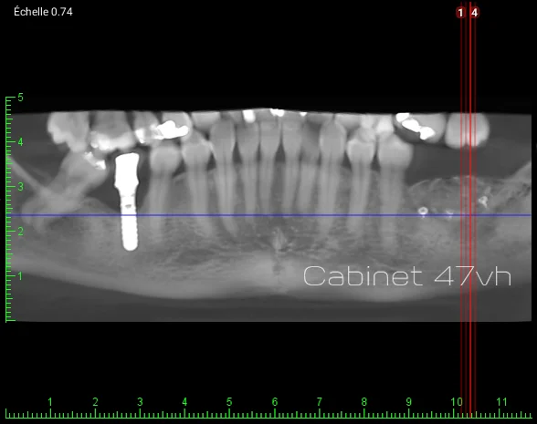
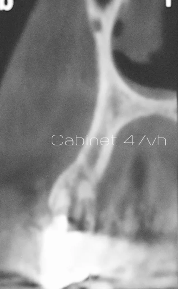
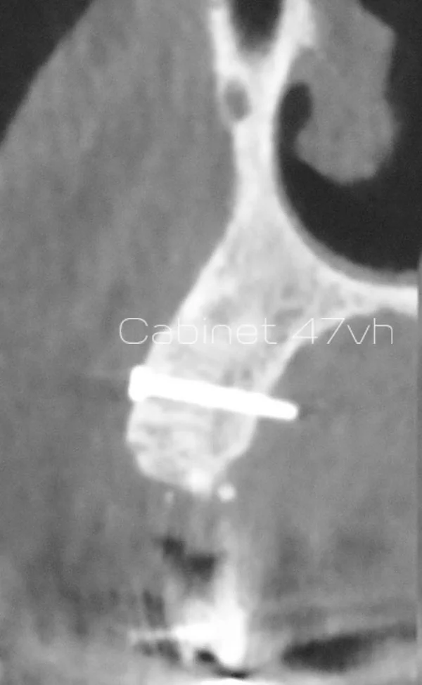
Bone grafting makes it possible to increase deficient bone volume by transplanting a bone graft to the area where additional bone is needed.
For autogenous bone grafts, bone is harvested directly from the patient, usually from another area of the jaw. For allogeneic grafts, a carefully prepared bone block from a human donor is used.
The bone fragment is adapted to the shape of the recipient site and then securely fixed with screws. After approximately four months, the grafted bone integrates with the jaw, allowing implant placement in sufficient bone volume.
Autogenous grafts provide highly reliable bone quality and quantity, but require surgery at a second site. Allogeneic grafts avoid this additional surgical site, although the bone quality and volume obtained may be less consistent.
Onlay autogenous graft
“
Dr Sylvain Mouraret’s opinion
In my practice, a bone graft is truly useful when the available bone volume does not allow the implant to be placed in a correct three-dimensional position. The goal is not simply to have “enough bone”, but to be able to position the implant according to the prosthetic plan, with an appropriate axis, depth, and implant emergence profile. When a graft is necessary, not performing it can compromise implant positioning and therefore the quality of the final prosthesis. Conversely, when bone volume is sufficient, we always favour the simplest and most predictable solution.
No. We recommend bone grafting only when the available bone volume is insufficient for predictable implant placement and long-term stability.
During the initial consultation, we perform a CBCT 3D scan to precisely assess your situation. In some cases, implants with adapted dimensions can be used to avoid this additional step.
Healing generally takes between 3 and 9 months, depending on the extent of the reconstruction.
For minor grafting procedures, healing is complete within 3 to 4 months. For more extensive reconstructions — such as a lateral sinus lift — healing may take up to 9 months. In favourable cases, grafting and implant placement can be performed during the same procedure, significantly reducing overall treatment time.
The procedure is performed under local anesthesia, so you will not feel any pain during the surgery.
After the procedure, moderate swelling and discomfort may occur for 5 to 7 days, and can be well managed with prescribed pain medication. Autogenous grafts may cause slightly more post-operative discomfort than biomaterial-based grafts.
The cost depends directly on the type and extent of bone reconstruction required.
A simple graft combined with implant placement is less expensive than complex vertical augmentation or a sinus lift. A detailed, personalised treatment plan is provided during the consultation after radiological assessment. We also offer interest-free payment options to facilitate access to treatment.
Some procedures may benefit from partial reimbursement depending on the clinical context.
Grafts performed as part of periodontal treatment may be partially covered by the National Health Insurance system. However, grafts performed in implant dentistry are usually not covered, although private insurance may contribute depending on your policy. We will explain the applicable coverage during your consultation.
Risks are rare when the procedure is performed by an experienced specialist.
They may include post-operative infection, partial failure of bone integration, or complications at the donor site in the case of autogenous grafts. We use only scientifically validated biomaterials to minimise these risks.
A sinus lift is a specific type of bone graft used exclusively in the posterior region of the upper jaw (maxilla).
It is performed when the bone height beneath the maxillary sinus is insufficient for implant placement. The sinus membrane is gently elevated to create space, which is then filled with a biomaterial. It is one of several bone augmentation techniques we routinely perform at the clinic.